


What’s that hand image?
PM&R News • November 13th, 2017
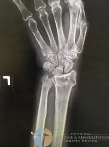
This image is demonstrating both a distal radius fracture and a distal ulnar fracture. Distal radius fractures are the most common type of orthopedic fracture and if the mechanism is high energy the ulnar can be involved as well. When the injury is this significant, the patient typically needs surgery to reduce and fix the fracture. Nonoperative indications of distal radius fractures are if the fracture is extra articular, if there is <5mm radial shortening, or if dorsal angulation is less than 5 degrees. A common complication of this injury is the development of carpal tunnel syndrome due to the space getting narrowed with the altered anatomy. Isolated distal ulnar fractures are less common and if the provider thinks they have an isolated distal ulnar fracture, one should take a second look at the x-ray to look for a radial fracture.
Review Question
Which tendon rupture is a common complication of distal radial fractures?Answer
EPL tendon ruptures are commonly seen in distal radial fractures, specifically nondisplaced fractures. When this occurs, it commonly is treated with transfer of extensor indicis proprius to EPL.
