


Multiple Myeloma
PM&R News • August 2021
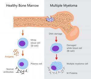
Anatomy
- Blood: Immunoglobulin-producing Plasma cells (derived from B cells; a lymphocyte sub-type)
Pathophysiology
- Plasma cells: neoplastic proliferation → monoclonal immunoglobulin
- Proliferation in the bone marrow = skeletal destruction
Epidemiology
- U.S.: 1 – 2% of all cancers
- > 17% of hematologic malignancies
- Annual: 4-5/100,000
- Worldwide: 160,000 cases
- 106,000 deaths/year
- Risk factors:
- Ethnicity:
- African Americans 2-3x > Caucasians
- Lowest risk: Asians from Japan, Mexicans
- Increased incidence: Males > females (1.4:1), ↑ BMI, older age (avg: 66 y/o), 1st degree relative (3.7x), variation of the 3p22.1 or 7p15.3 genetic loci
- Ethnicity:
Presentation/ Clinical Manifestations
- Anemia: 73% at diagnosis; 97% at some time during disease course
- fatigue/pallor
- Bone pain: 60%
- Greatest at the back/chest secondary to movement (none at night); vertebral collapse, rib Plasmacytomas (tumor in ST or skeleton)
- Renal insufficiency: ~50%
- Increased serum creatinine concentration
- 2 main causes: light chain cast nephropathy (aka: myeloma kidney) and hypercalcemia
- Increased serum creatinine concentration
- Hypercalcemia: 13%
- Cord compression: 5%
- secondary to vertebral fx or extramedullary plasmacytoma
- s/s: back pain with weakness and paresthesias; B/B disfunction or incontinence.
The following percents: a retrospective analysis of 1027 sequential patients diagnosed with MM at a single institution:
- Anemia – 73%
- Bone pain – 58%
- Elevated creatinine – 48%
- Fatigue/generalized weakness – 32%
- Hypercalcemia – 28%
- Weight loss – 24%, (50% lost ≥9 kg/19.84 lbs)
- < 5%: paresthesias (5%), hepatomegaly (4%), splenomegaly (1%), lymphadenopathy (1%), and fever (0.7%).
- Acute renal failure or nephrotic syndrome (rare)
Diagnosis
- Imaging (preferred: cross-sectional due to increased sensitivity): lytic lesions present on bone (MRI can detect lesions not yet lytic)
- SPEP (protein electrophoresis of the serum) or UPEP (urine protein electrophoresis) combined with immunofixation (serum or urine): presence of a monoclonal protein (97%)
- total serum protein concentration: increased — typically normal in those with light-chain myeloma (detection via: serum FLC (free light chain), UPEP and urine immunofixation)
- Bone marrow bx: clonal plasma cells >/=10% (96% pts; average typically around 50%)
- If >60%, 80% progress to end-organ damage in 2 years
- Hypercalcemia: >11 mg/dL (>2.75 mmol/liter).
- Histopathology specimen: (+) plasmacytoma
- Immunophenotype: express CD56 (~70%)
- Free light chain assay: abnormal (90%)
-
Definitive dx:
Clonal bone marrow plasma cells ≥10 percent or bony or soft tissue plasmacytoma (demonstrated via biopsy) + one of the following:
- (CRAB: calcium level, renal insufficiency, anemia, and bone lesions)
“Presence of a biomarker associated with near inevitable progression to end-organ damage”
Treatment
- autologous hematopoietic cell transplantation (HCT): prolong event-free & overall survival
- Chemo:
- HCT eligible: triplet regimen, 3-4 mos
- Non-eligible: triplet regimen, 8-12 cycles; maintenance therapy until progression
- Frail: doublet therapy until progression
- Both require maintenance therapies
Patient Education
- No cure: Tx aimed at alleviating symptoms, inducing remission, extending life.
- Increased risk of infection secondary to:
- Hypoventilation
- Decreased lymphocytes, plasma cell suppression, and hypogammaglobulinemia
References:
- https://www.uptodate.com/contents/multiple-myeloma-clinical-features-laboratory-manifestations-and-diagnosis?search=multiple%20myeloma&source=search_result&selectedTitle=1~150&usage_type=default&display_rank=1
- https://www.uptodate.com/contents/multiple-myeloma-symptoms-diagnosis-and-staging-beyond-the-basics?search=multiple%20myeloma&topicRef=6643&source=see_link
Review Question
Which of the following clinical manifestations is most common at diagnosis? Bone pain, Anemia, hypercalcemia, renal insufficiency, or cord compression?Answer
Anemia (fatigue, pallor, pica, etc…) → 73% at diagnosis; 97% at some time during disease course
