


Avascular Necrosis
PM&R News • December 2020
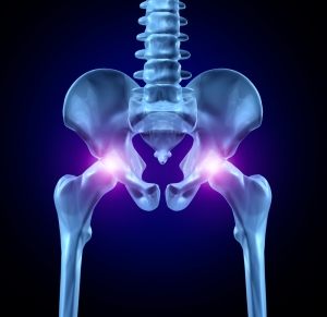
A 41-year-old male presented to a chiropractor’s office for ongoing right sided low back, hip and knee pain for the past six months following jumping off a two meter high roof and landing on his feet. He had immigrated to Canada from abroad two months prior to consulting us. The patient had an antalgic limp and walked with the help of a cane. He complained of intermittent pain radiating into his right groin and anteriomedial thigh region. He stated that his symptoms were aggravated by walking and stair climbing. His pain was relieved by sitting and resting. The patient did not report numbness or paresthesias in his lower extremities. There was no bowel and bladder dysfunction. The patient did not complain of any night sweats, fever or chills.
Description
- Other common names: Osteonecrosis, aseptic necrosis, atraumatic necrosis, or ischemic necrosis.
- Pathologic: Typically 2/2 direct bony or vascular injury
- Etiology: MC due to mechanical failure
- Most common: “ASEPTIC”
- A – Alcohol/AIDS
- S – Steroids, sickle cell disease, SLE
- E – Erlenmeyer flask (Gaucher disease)
- P – Pancreatitis
- T – Trauma
- I – Idiopathic, infection
- C – Caisson’s disease
- Pediatric types:
- idiopathic osteonecrosis of the femoral head (Legg-Calvé-Perthes disease)
- osteonecrosis 2/2 a slipped capital femoral epiphysis (SCFE)
- Most common: “ASEPTIC”
Anatomy
- MC sites of occurrence: femoral head (others: humeral head, talus, scaphoid. Any bone, however, can have this occur.
- Arterial circulation
- Femoral neck: medial femoral circumflex artery (ascending branch)
Pathophysiology: Arterial damage → ↓ perfusion → ischemia → bone/marrow death → Joint death (months/years)
Epidemiology
- U.S.: 10,000-20,000 / year (Hip)
- 35-50 y/o
- MC in males > females
- Risk factors
- traumatic injury (affects arteries)
- Medications/Conditions that alter perfusion
- Ie: glucocorticoids, bisphosphonates, sickle cell disease, SLE, Gaucher disease [GD], thrombophilia, decompression disease (ie: dysbarism or caisson disease; nitrogen bubbles occlude arterioles)
- Bone Marrow toxic treatments: radiation therapy (osteoradionecrosis)
- Post-transplation (50-20% of renal transplants)
- Alcohol
- Tobacco use
- Acute lymphoblastic leukemia (2.3% development w/in 5 years presumably d/t glucocorticoid tx)
- HIV infxn (45-100x greater)
- Postop knee arthroscopy
- Spontaneous osteonecrosis of the knee (SPONK)
Differential Diagnoses
- Bone marrow edema syndrome, BMES (aka: transient osteopenia of the hip, TOH)
- MRI: decreased T1 signal; increased intensity on T2 images
- Subchondral insufficiency fx → MRI: low signal in subchondral region on T1/2 imaging
- Idiopathic transient osteoporosis of the hip: MRI: femoral neck and metaphysis edema
- Osteonecrosis during pregnancy
- Peripheral arterial disease (PAD)
Presentation/ Clinical Manifestations
- Asymptomatic
- Symptoms
- Pain +/- activity (MC presenting symptom): Groin > thigh > buttocks (if femur)
- With weightbearing and/or motion
- Rest pain: 2/3
- Nocturnal: 1/3
- “Many have bilateral involvement at the time of diagnosis”
- Physical Exam findings: Non-specific
- ↓ ROM (esp. forced IR and abduction à joint compaction)
- Limp (late in course of disease)
- Normal pedal pulse exam (unless simultaneous vascular issues)
- Pain +/- activity (MC presenting symptom): Groin > thigh > buttocks (if femur)
Diagnosis
- Imaging
- Initial: plain radiograph (XR)
- Negative, but highly suspicious still; perform MRI
- Not recommended: radionuclide bone scanning
- Initial: plain radiograph (XR)
- Definitive Diagnosis: clinical (based on hx and XR or MRI findings)
- Gold standard: MRI without contrast
- ARCO Staging (Association Research Circulation Osseous):
- I: normal XR/abnormal MRI
- II: XR — sclerosis, osteolysis or focal porosis // No crescent sign
- III: subchondral fx, or fx in the necrotic portion, or flattening of the femral head
- A: femoral head depression < 2 mm
- B: femoral head depression >2 mm
- IV: OA, ↓ joint space, acetabular changes
Treatment
- Non-pharmacologic (Investigational; unreliable improvement)
- Bisphosphonates (asymptomatic, <15% or >30% of the femoral head, & no collapse)
- Pharmacologic (Investigational; unreliable improvement)
- Statin therapy
- Anticoagulation
- Vasodilators
- Others (Investigational; unreliable improvement)
- extracorporeal shock wave therapy (ESWT)
- electrical/electromagnetic stimulation
- hyperbaric oxygen
- Surgical
- Femoral head core decompression +/- stem cell injection asymptomatic + 15-
- Symptomatic + ARCO Stage 1 or 2 30% involvement
- Vascularized free-fibula grafting
- ARCO Stage IIIA
- Total hip resurfacing
- Total hip arthroplasty (THA)
- ARCO Stage IIIB or IV
- Femoral head core decompression +/- stem cell injection asymptomatic + 15-
Complications:Femoral collapse (risk determined by modified Kerboul à necrotic angles <190º, 190-240 º, >240 º correlated with mild, mod, and high risks., respectively).
Prognosis: <10% collapsed if small/medial lesions (hip)
References:
- https://www.uptodate.com/contents/clinical-manifestations-and-diagnosis-of-osteonecrosis-avascular-necrosis-of-bone#H878671233
- Vignette
- https://www.uptodate.com/contents/treatment-of-nontraumatic-hip-osteonecrosis-avascular-necrosis-of-the-femoral-head-in-adults?topicRef=122049&source=see_link#H18899908
Review Question
What is the gold standard for diagnosis of Avascular Necrosis?Answer
MRI without contrast
